Website Banner (2).png

Established since 1999, X-Ray Newstead has been providing high quality, personalised radiology services in Launceston. 


With a full accreditation with Quality, Innovation, Performance (QIP) X-Ray Newstead is fully equipped for all general practice, dental x-rays (including Cone beam CT) and ultrasound services.

Launceston is home to the first x-ray photographs in the southern hemisphere, with Frank Styant Browne, creating the first x-ray photograph in 1896. Since then, Launceston has been a pioneer of the latest in x-ray technology.


We accept any and all referrals.

No matter where it is written for.


Our Services

At X-Ray Newstead, we are committed to providing our patients with the highest quality information, diagnostic imaging, and care. Below is a sample of what we offer:

Dental

X-Ray Newstead provides a range of dental imaging.

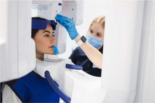
The most common dental examination we offer is an OPG (Orthopantomogram). The OPG provides a panoramic view of the mouth, teeth and jaw, and is particularly useful in showing the position and growth of teeth.

A Cephlastat is a side-on (lateral) view of the face. It allows for precise measurements to be made and enables the relationship between the top and bottom jaw to be seen. Cephlastats are also useful in planning orthodontic treatments.

We also offer Cone Beam CT imaging. This type of imaging provides high resolution 3D images and allows teeth and facial bones to be viewed in any plane. Cone Beam CT imaging is useful for examining impacted teeth, planning dental implants and assessing pathology and trauma.

General

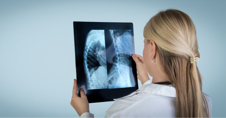
General radiography (or x-ray) refers to the use of x-rays to create static 2D images of different parts of the body.

An image is produced when a small amount of radiation passes through a patient’s body and is recorded on a film or detector on the other side of the body.

X-ray is the most commonly performed examination and is very good for investigating bones as well as soft tissue such as the chest, lungs and abdomen.

Unless a more complex investigation is required, plain x-rays are generally very quick and don’t cause any discomfort to the patient.

Ultrasound

Ultrasound is a widely used and effective tool for examining a variety of body areas and organs.

It does not produce any ionizing radiation and works by sending high frequency sound pulses into the body through a handheld probe or transducer.

The echoes or soundwaves that are bounced back to the transducer create high resolution images of the body part being scanned.

Ultrasound can be used for a wide variety of diagnostic purposes, including pregnancy scanning and imaging of the liver, kidneys, muscles, tendons, veins, arteries, neck, and breast.

Some ultrasound examinations require pre-scan preparation.  If you have any queries, please refer to our patient preparation sheets or simply phone one of our clinics.